O-pH: Optical pH Monitor to Measure Oral Biofilm Acidity and Assist in Enamel Health Monitoring

Manuja Sharma, Lauren K. Lee, Matthew D. Carson, David S. Park, Se W. An, Micah G. Bovenkamp, Jess J. Cayetano, Ian A. Berude, Leonard Y. Nelson, Zheng Xu, Alireza Sadr, Shwetak Patel, Eric J. Seibel
PDF
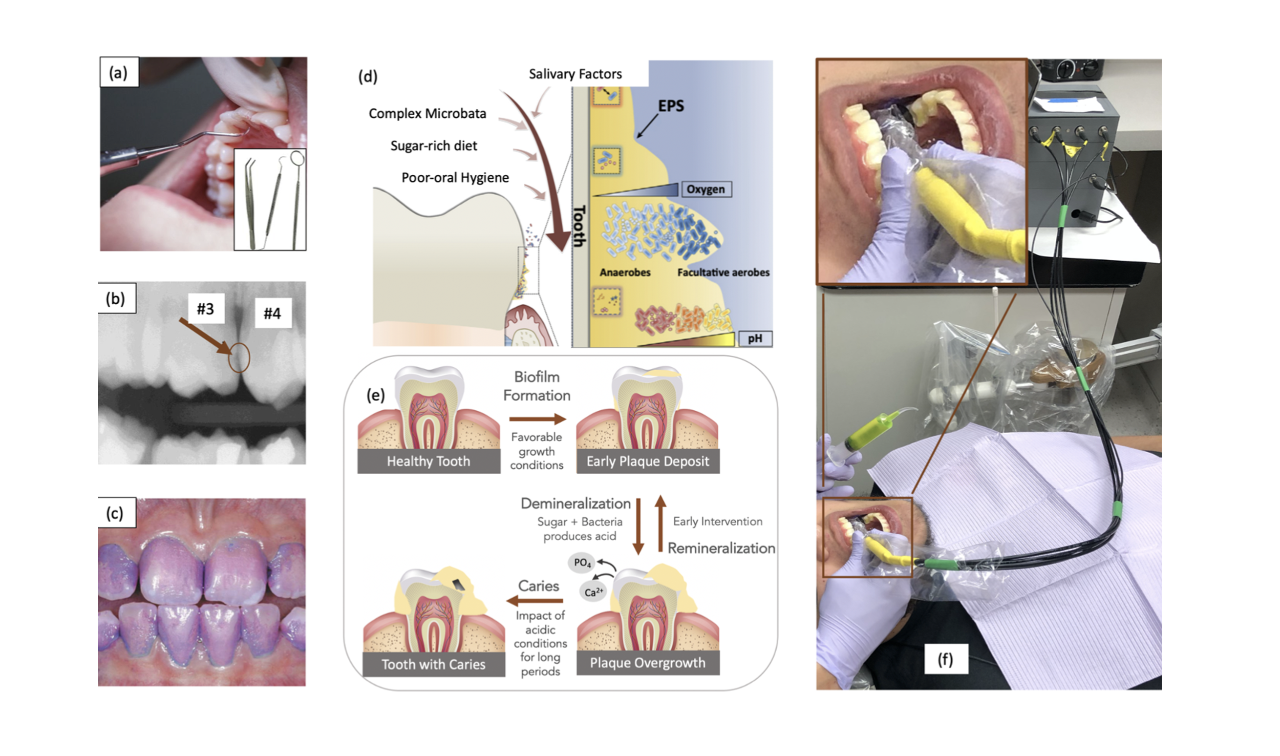
(a) Visual assessment using dental tools - gold standard for early occlusal caries. Inset figure shows different kinds of probing instruments used by dentists (b) Bitewing X-ray with an interproximal lesion between teeth 3 and 4 - gold standard for early interproximal caries (c) Patient’s mouth after using a biofilm disclosing agent to see dental biofilm coverage (d) Biofilm micro-environment: pH level is lower moving from surface to enamel. Extracellular Polymeric Substance (EPS) composition and characteristic is shown in the inset figure. (e) Caries formation (f) O-pH in operation at a dental clinic with an inset figure showing a closer look of the device inside the mouth. The tip of the probe used to transmit and collect light is hovering over the occlusal surface of the subject.

Abstract

Objective: Bacteria in the oral biofilm produce acid after consumption of carbohydrates which if left unmonitored leads to caries formation. We present O-pH, a device that ca measure oral biofilm acidity and provide quantitative feedback to assist in oral health monitoring. Method: O-pH utilizes a ratiometric pH sensing method by capturing fluorescence of Sodium Fluorescein, an FDA approved chemical dye. The device was calibrated to a lab pH meter using buffered fluorescein solution with a correlation coefficient of 0.97. The calibration was further verified in vitro on additional buffered solution, artificial, and extracted teeth. An in vivo study on 30 pediatric subjects was performed to measure pH before (rest pH) and after a sugar rinse (drop pH), and the resultant difference in pH (diff pH) was calculated. The study enrolled subjects with low (Post-Cleaning) and heavy (Pre-Cleaning) biofilm load, having both unhealthy/healthy surfaces. Further, we modified point-based O-pH to an image-based device using a multimode-scanning fiber endoscope (mm-SFE) and tested in vivo on one subject. Results and conclusion: We found significant difference between Post-Cleaning and Pre-Cleaning group using drop pH and diff pH. Additionally, in Pre-Cleaning group, the rest and drop pH is lower at the caries surfaces compared to healthy surfaces. Similar trend was not noticed in the Post-Cleaning group. mm-SFE pH scope recorded image-based pH heatmap of a subject with an average average diff pH of 1.5. Significance: This work builds an optical pH prototype and presents a pioneering study for non-invasively measuring pH of oral biofilm clinically.